No matter what time of day, virtually no one is getting nervous about running a cranial CT scan (CCT. It's a routine exam that's one of the first things you learn and one of the most frequently performed. The standard dictates that the patient lies on their back with their arms next to their body, and after a few minutes, the images are ready. Basically, not a big challenge.
If only it were always so "simple". There are some patients who simply won't or can't lie still. For example, patients with suspected stroke or dementia. Although stabilization belts can help in these situations, they are not always effective. Accordingly, it is important in practice to be able to perform examinations in a way that is different from the routine.
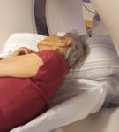
Patients with symptoms of vertigo, headaches, combined head and back pain or patients with dementia often prefer to lie on their side with their legs bent. Then, this is an effective, alternative examination procedure (see picture to the right).
If a blanket is then placed over the patient, they usually lie still in this position and good quality images can be taken.
Attention should be paid to the following points:
- Slightly secure the patient on the table by means of a stabilization belt.
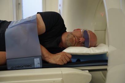
- Use radiation protection for the eyes (see picture below left, patient in lateral position with belt fastening and lens protection; ProBelt and PearlFit positioning aids)
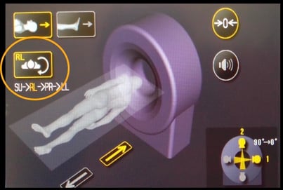
- Which side is the patient lying on? Enter patient position on the CT (see picture below right, example of patient position on Canon CT).
- Deactivate dose modulation
- Prepare reconstructions
The dose modulation should be deactivated in any case, as depending on the manufacturer it can only work correctly ventrally and dorsally, which relates to the lateral side of the patient's head in this case.
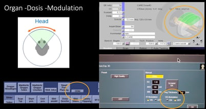
Activated dose modulation can lead to overexposure (too much radiation) of one and underexposure (too little radiation) of the other side. Furthermore, usually dose modulation is used to protect the lenses of the eye from radiation; however, they are not in the beam corridor in this case, compared to a normal CCT. (see picture to the right, Deactivating dose modulation) At Siemens, a CCT protocol without X-Care is to be used.
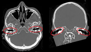
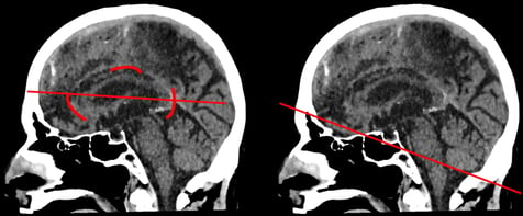
For the final reconstructions, look for the internal auditory canals on the axial and coronal images (see picture below left) and the anterior and posterior knees of the collum callosum on the sagittal image (see picture below right). Alternatively, tilt along the base of the skull to create correct reconstructions in the soft tissues and, if necessary, in the bone window.
Lateral positioning of the patient during CCTs may take some courage. However, it is a relief for patients and radiology staff in certain situations as difficulties in positioning the patient can be mitigated.
Credits to: Dorina Petersen for providing this content https://www.dorina-petersen.de
_ _ _ _ _ _ _ _ _ _ _ _ _ _ _ _ _ _ _ _ _ _ _ _ _ _ _ _ _ _ _ _ _ _ _ _ _ _ _ _ _ _ _ _ _ _ _ _ _ _ _ _
ABOUT PEARL TECHNOLOGY
Pearl Technology AG, based in Schlieren, offers innovative solutions for the placement, positioning and immobilization of patients in radiology and radiotherapy. The products are manufactured in Switzerland according to ISO norm 13485 and are characterized by simple handling, high patient comfort and excellent hygiene which guarantees smooth and safe examination processes.
Download further information on our patient positioning portfolio for radiology and nuclear medicine or contact us directly in order to receive further information on our patient positioning solutions.

