Correct patient positioning is a critical factor in the safety and success of imaging examinations. However, even experienced professionals can sometimes make mistakes when positioning patients, unintentionally creating risks for the patient. This blog explains two common mistakes in patient positioning and describes how to avoid them for a safe and effective examination.
ERROR 1: THE EDGE OF THE Positioning AId LIES ON THE POINT OF INTEREST (POI)
The "invisible" and hygienic positioning aid does not exist. And so, if the positioning aid is incorrectly positioned, it may be the case that it lies exactly on a fracture line. There is a possibility that it will cause a shadow on the image, which can affect the accuracy of the diagnosis. This occurs, for example, when the patient is lying off-centre on the positioning mat.
Examples of this are:
- Pelvic exam: Patient lies off-centre on the edge of the mattress.
- Supine Y-view: Wedge is placed under the patient's shoulder.

This problem can be significantly reduced if hard, dimensionally stable positioning aids are used, which allow patients to be placed in the best possible inclined position (for example with the ProFoam DS).
Advise: For the shoulder exam, the positioning wedge should be positioned in the lower thoracic spine area. This allows the shoulder to be exposed without a wedge and the patient lies much more stable in the oblique position.
ERROR 2: AVOID TAMPERING WITH THE positioning equipment
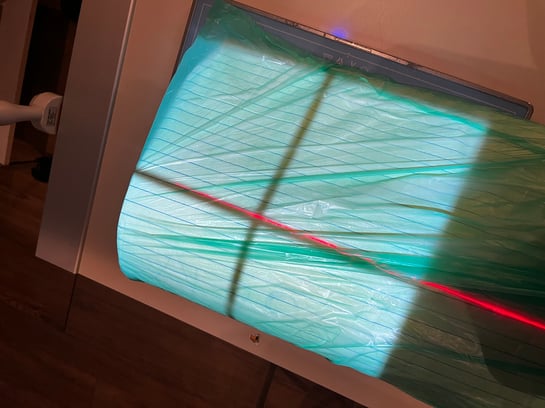
Radiology positioning aids should comply with hygiene standards and medical product regulations. However, the hygienic standard cannot be ensured by wrapping the positioning aids in a plastic bag (Fig. 1).
To get a better impression of this issue, an X-ray image of a wrapped knee wedge at 50kV is illustrated here. Clear artefacts are visible on the image, which were caused by the wrapping of the foam.
.jpeg?width=305&height=406&name=Image%20(1).jpeg)
As mentioned above, the "invisible" and at the same time hygienic positioning aid unfortunately does not exist. In order not to run any unnecessary hygiene risks, positioning aids sealed in PU foil (or comparable materials) from specialised manufacturers should be used (for example, ProFoam product family from Pearl Technology).
Although these types of positioning aids are also partially visible on the X-ray images, clear work procedures can be established and standards defined thanks to the always identical product properties. For example, the positioning aid is used outside the field of view or the placement of the edge can be cleverly chosen (compare, point 1 above).
More on this topic can also be found in this Blog.
CONCLUSION
By choosing suitable aids and using them correctly, errors in patient positioning can be avoided. This can reduce risks to the patient and ensure efficient and effective examination procedures.
Credits to: Michael Bünte, radiographer, for the preparation of the content (https://mtrnrw.de).
About PEARL TECHNOLOGY
Pearl Technology AG, based in Schlieren, offers innovative solutions for the placement, positioning and immobilization of patients in radiology and radiotherapy. The products are manufactured in Switzerland according to ISO norm 13485 and are characterized by simple handling, high patient comfort and excellent hygiene which guarantees smooth and safe examination processes.
Download further information on our patient positioning portfolio for radiology and nuclear medicine or contact us directly in order to receive further information on our patient positioning solutions.
