Positioning the shoulder requires a precise alignment of the patient in order to achieve optimal imaging results. Particular precision is required if the patient has to be X-rayed lying down. If the shoulder is injured in an accident, it is often the case that patients are unable to sit or stand. In order to achieve a very good result despite this, a conscious approach to positioning is required. When applied correctly, the diagnostic significance of the supine shoulder image is comparable to the version when the patient is standing or sitting.
Below are tips with background information and advice on adequate positioning with the 45° wedge.
THE RIGIDITY MAKES THE DIFFERENCE
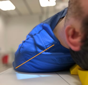
What criteria should large 45° wedges fulfil? Is it the case that a wedge that is too soft will not manage to hold the patient in the required position? In a test, we compare two different wedges. A conventional, coated foam (yellow version shown here) and a ProFoam Wedge 60x25x12 DS from Pearl Technology, whereas the conventional foam is slightly softer than the ProFoam wedge.
Despite the different degrees of rigidity, both wedges are able to hold the test subject (184 cm and 98 kg) in position for the ap and Y-view. However, the harder wedge yields less and the patient does not sink in. In comparison, the subject also noticed that the blue wedges were slightly harder and that he was lying more firmly as a result, but also that the blue wedge slipped slightly more on the detector. However, this is easily prevented by using a sandbag.
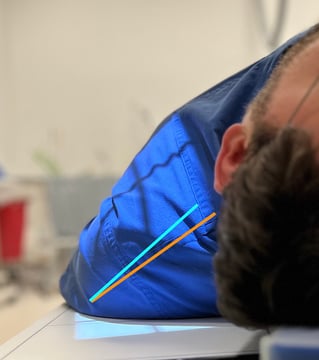
The images below show that in the AP view, the subject settles slightly on the yellow wedge. On the blue wedge, the patient does not sink in at all, which makes the recording ideal.
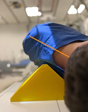
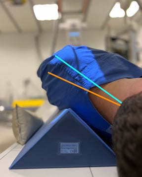
The difference is even more pronounced in the Y-view. The yellow wedge yields significantly more than the blue ProFoam Wedge 45°. The ProFoam Wedge 45° wedge is rigid enough to hold the patient in the correct position and is therefore ideal for this setting. The pictures below show a Y-view AP in supine position - a suitable backup option when patients cannot stand or sit.
IMPORTANT NOTE ON THE IDEAL PLACEMENT OF THE 45° WEDGE
The ideal placement of the 45-degree wedge is important to avoid the interference of edges of the positioning aid on X-ray images and to ensure that the diagnostic value is not impaired by line-shaped structures that could look like fracture lines.
Three options for positioning the wedges are illustrated and assessed below.
Option 1 - faulty


The patient is lying stable, but the edge of the positioning aids can be recognised on the image and may impair diagnosis.
Option 2 - correct


The patient is lying stable and the edge of the positioning aids cannot be seen on the image and therefore does not impair the image quality.
Option 3 - correct


The patient is lying stable, the edge of the positioning aid is not visible on the image and therefore does not impair the image quality. Adjusting the X-ray marker is slightly more difficult.
Conclusion
The choice of suitable positioning aids has a direct impact on workflow and the quality of the examination results. More rigid wedges are preferable for shoulder AP and Y-imaging in supine position, as the patient remains in the desired 45° position. A sandbag can help to additionally stabilise the position. Skilful and careful use of the positioning aids ensures that no edges are visible on the images and ensures satisfactory results.
Author: Agata Epler, Independent X-Ray Instructor at Alles Einstellungssache